Plant Anatomy, Again with Dr. Jim Mauseth
Update: 2024-10-16
Description
If the ads are bumming you out, keep in mind that ad-free episodes of the podcast are available at :
www.patreon.com/crimepaysbutbotanydoesnt
Did you know that the distal ends and tips of roots are the only parts doing any absorption? What the hell are cortical bundles and why did cacti evolve them? How can cactus roots grow so quickly after a rain and what do we mean by "root spurs"? How does the South American parasitic plant Tristerix aphylla behave like a fungus when it grows inside its host plant? And if you still don't understand what the hell Parenchyma is, here's your chance for a refresher.
Dr. Jim Mauseth taught plant anatomy and botany for 30 years at UT Austin and literally wrote a textbook on the subject. He's also written a few other books and over a hundred research papers studying the anatomy of plants with an emphasis on cacti, and has traveled to South America and Mexico studying the family on numerous occasions. In this episode we go deep on plant tissues, plant cells, cellular components, plasmodesmata, cell membranes and how the a plant is technically only one single cell when you really get down to it...
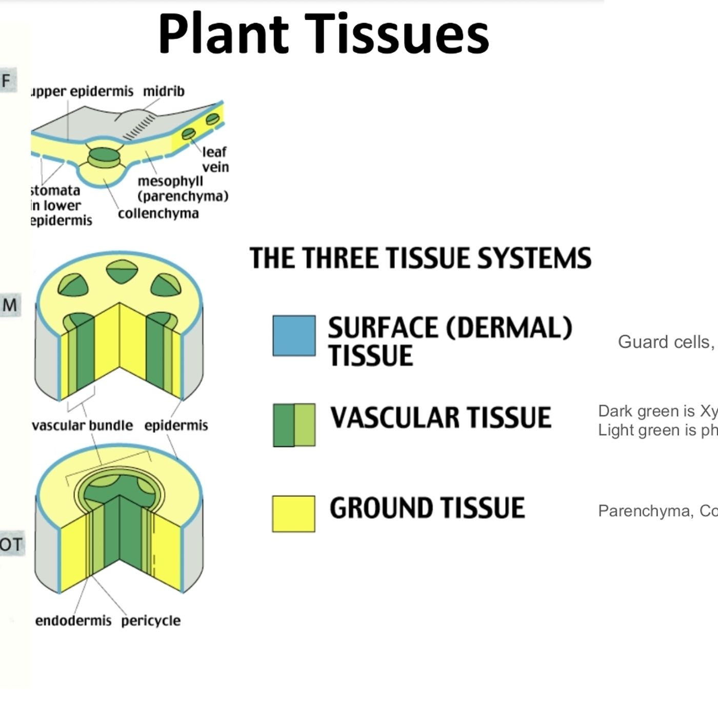
A reminder that the previous podcast episode on plant tissues covers some of the terminology in this episode, such as the 3 main tissue types : epidermal tissues, ground tissues (parenchyma, collenchyma, sclerenchyma) and vascular tissue (xylem and phloem). I highly suggest listening to that episode first or at least pausing the podcast if you're unclear about some of the terminology. Remember that tracheids and vessel elements apply only to xylem (which only moves water) and "sieve tubes", "companion cells" and "sieve plates" apply only to phloem (which only moves sugars and photosynthates).
The 3 ground tissues are : parenchyma (primary walls only, large intercellular spaces, alive at maturity), collenchyma (only produces primary cell walls with thickened and re-inforced corners, alive at maturity), sclerenchyma (primary and secondary cell walls, dead at maturity).
Thumbnail photo shows the incredibly thick cuticle of Ariocarpus, with epidermis below and hypodermis below that, marked with arrows. Vertical hole on the right side is the stomatal opening in the cuticle
Comments
Top Podcasts
The Best New Comedy Podcast Right Now – June 2024The Best News Podcast Right Now – June 2024The Best New Business Podcast Right Now – June 2024The Best New Sports Podcast Right Now – June 2024The Best New True Crime Podcast Right Now – June 2024The Best New Joe Rogan Experience Podcast Right Now – June 20The Best New Dan Bongino Show Podcast Right Now – June 20The Best New Mark Levin Podcast – June 2024
In Channel


