The Trigeminal Nerve Block and Cancer (Ultrasound and Flouroscopic Approaches)
Update: 2024-07-19
Description
PainExam Show Notes: Mandibular Division of the Trigeminal Nerve Block with Dr. David Rosenblum
Introduction
- Host: Dr. David Rosenblum
- Topic: Mandibular Division of the Trigeminal Nerve Block for Cancer Pain Management
- Techniques: Ultrasound and Fluoroscopic Guidance
Overview
- Purpose: Alleviate chronic facial pain, specifically in cancer patients suffering from trigeminal neuralgia or other related conditions.
- Focus: Detailed discussion on the anatomy, clinical presentation, and procedural techniques for effective nerve block.
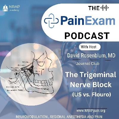
Anatomy of the Mandibular Nerve
- Origin: Mandibular nerve is a branch of the trigeminal nerve (cranial nerve V).
- Pathway: Exits the middle cranial fossa through the foramen ovale and descends between the lateral and medial pterygoid muscles.
- Sensory Innervation:
- Anterior two-thirds of the tongue
- Teeth and mucosa of the mandible
- Skin of the chin and lower lip
- Skin over the mandible (excluding the mandibular angle)
- Tragus and anterior part of the ear
- Posterior part of the temporalis muscle up to the scalp
Ultrasound-Guided Technique
- Patient Positioning:
- Patient lies on their side with the affected side facing upward.
- Transducer Selection:
- Curvilinear transducer preferred for deeper structures.
- Transducer Placement:
- Place distal and parallel to the zygomatic arch to bridge the coronoid and condylar processes.
- Anatomical Landmarks:
- Identify the lateral pterygoid muscle and plate.
- Use power Doppler to locate the sphenoid palatine artery.
- Needle Trajectory:
- Introduce the needle using an out-of-plane approach to target the pterygopalatine fossa (anterior to the lateral pterygoid plate).
- For the mandibular nerve block, target the area posterior to the lateral pterygoid plate between the medial and lateral pterygoid muscles.
- Electrostimulation (Optional):
- Utilize a 22G, 10 cm insulated short beveled needle connected to a peripheral nerve simulator.
- Position confirmed by motor response from the temporalis and masseter muscles.
Fluoroscopic-Guided Technique
- Patient Positioning:
- Similar to ultrasound guidance, patient lies on their side with the affected side facing upward.
- C-arm Positioning:
- Position the C-arm to visualize the foramen ovale.
- Needle Insertion:
- Insert the needle under fluoroscopic guidance towards the foramen ovale.
- Contrast Injection:
- Confirm needle placement with contrast injection.
- Anesthetic Administration:
- Administer local anesthetic and/or neurolytic agents.
Clinical Symptoms and Diagnosis
- Symptoms:
- Unilateral sharp, stabbing, or burning pain in the mandibular nerve distribution.
- Pain triggered by activities such as eating, talking, washing the face, or cleaning the teeth.
- Diagnostic Imaging:
- MRI or CT scans to identify causes like vascular compression, mass lesions, or fractures.
Complications and Considerations
- Potential Complications:
- Bleeding, hematoma, infection, and hypersensitivity reaction to the injectate.
- Serious complications from neurolytic agents like permanent sensory deficit and tissue necrosis.
- Alternative Treatments:
- PNS? Radiofrequency or cryoablation for recalcitrant cases.
Conclusion
- Efficacy: Ultrasound and fluoroscopic guidance provide precise targeting of the affected nerves, minimizing collateral damage.
- Safety: Routine use of power Doppler imaging to avoid injury to surrounding vessels.
- Recommendation: Consider these techniques for patients unresponsive to oral medications or unsuitable for surgery.
These show notes provide a comprehensive overview of the discussion, highlighting key points on the anatomy, technique, and clinical considerations for mandibular nerve blocks in cancer patients.
Comments
In Channel

